Myositis Ossificans Radiology, Soft Tissue Calcifications
Myositis ossificans radiology Indeed lately is being hunted by consumers around us, maybe one of you. Individuals are now accustomed to using the internet in gadgets to view image and video information for inspiration, and according to the name of this post I will discuss about Myositis Ossificans Radiology.
- Heterotopic Ossification Wikipedia
- Cureus Atypical Presentation Of Fibrodysplasia Ossificans Progressiva A Case Report And Review Of Literature
- Myositis Ossificans Radsource
- Myositis Ossificans Radsource
- Myositis Ossificans Progressiva A Clinico Radiological Evaluation Case Report With Brief Review Of Literature Rathee N Gupta Pk Gupta K Garg G J Orthop Allied Sci
- Https Encrypted Tbn0 Gstatic Com Images Q Tbn And9gcsneywrg987xc0x2gltgarpmjjeecmjkiqsmft1wj80lefj Abg Usqp Cau
Find, Read, And Discover Myositis Ossificans Radiology, Such Us:
- Soft Tissue Calcifications A Pictorial Essay
- Myositis Ossificans Radiology Case Radiopaedia Org
- 99 Heterotopic Ossification Myositis Ossificans Radiology Key
- Myositis Ossificans Radiology Case Radiopaedia Org
- Cureus Atypical Presentation Of Fibrodysplasia Ossificans Progressiva A Case Report And Review Of Literature
If you are searching for Meijer Thanksgiving 2020 Sale you've reached the ideal place. We ve got 104 graphics about meijer thanksgiving 2020 sale including images, photos, pictures, backgrounds, and much more. In these web page, we also have variety of images available. Such as png, jpg, animated gifs, pic art, logo, black and white, transparent, etc.

Figure 7 From The Imaging Of Myositis Ossificans Semantic Scholar Meijer Thanksgiving 2020 Sale

Myositis Ossificans Nejm Meijer Thanksgiving 2020 Sale

Myositis Ossificans Imaging Keys To Successful Diagnosis Lacout A Jarraya M Marcy Py Thariat J Carlier Ry Indian J Radiol Imaging Meijer Thanksgiving 2020 Sale

Myositis Ossificans Imaging Keys To Successful Diagnosis Lacout A Jarraya M Marcy Py Thariat J Carlier Ry Indian J Radiol Imaging Meijer Thanksgiving 2020 Sale
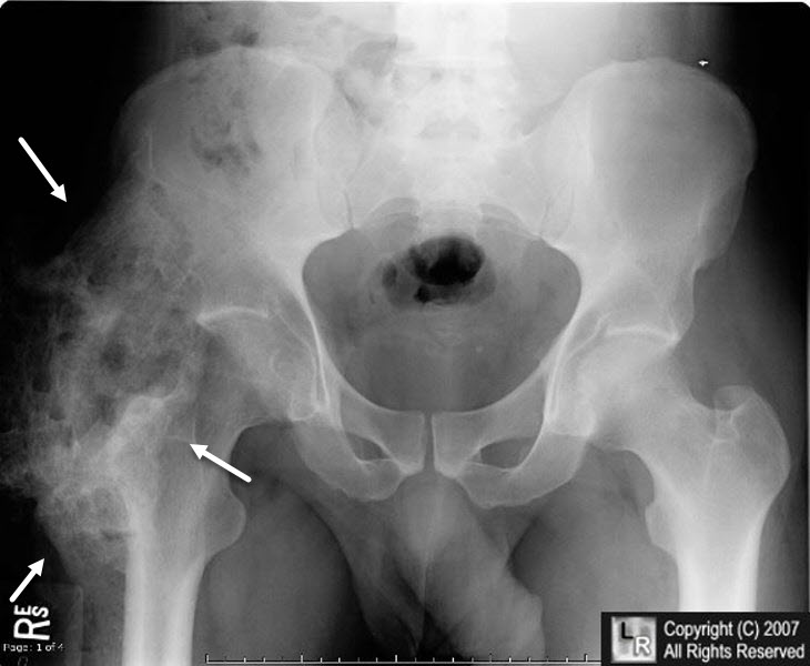
Learningradiology Heterotopic Ossification Ho Meijer Thanksgiving 2020 Sale
Http Pdf Posterng Netkey At Download Index Php Module Get Pdf By Id Poster Id 102318 Meijer Thanksgiving 2020 Sale
Myositides can be generally categorized by etiology as follows 1.
Meijer thanksgiving 2020 sale. It progresses through characteristic stages both histologically and on imaging studies. Mr imaging features particularly after injection of gadopentetate dimeglumine mimicked those of an inflammatory mass or neoplasm. Patients with available mr images were chosen from a group of 326 cases in our radiologic archives of histologically proved and radiologically correlated myositis ossificans.
Complete maturation of the mass occurs in 612 months. Progressive and diffused ossification of tendons ligaments and connective tissue around muscles micro clinodactyly. Keys to the diagnosis are the circumscribed calcification on x ray location that is prone to trauma in the an.
Mr imaging has unique advantages for diagnosis of early mo without calcification or ossification. Mri findings of early myositis ossificans without calcification or ossification. It actually looks more sinister on mri than x ray or us.
The ossification appears to be contiguous with the shaft of the femur with no intervening fascial plane. The final diagnosis was myositis ossificans mo. We reviewed retrospectively the mr images of eight histologically proved cases of myositis ossificans and correlated the mr appearance with the histologic findings as well as with other radiologic studies.
The lesions were excised in three patients and the images were correlated with histologic findings. There are some conditions that are related to or share a similar name to myositis ossificans 1. Myositis ossificans is a self limiting typically post traumatic cause of bone producing intramuscular lesions.
The ct scan demonstrates a curvilinear area of ossification lying adjacent to the lateral aspect of the mid left femoral shaft. Myositis ossificans thigh. The early stage which occurs within 4 weeks.
Mri can delineate the extent of the tumor and. This is an important diagnosis to make radiologically so as to avoid biopsy. Painful enlarging mass with intense inflammation of the surrounding tissues.
The intermediate stage seen at 48 weeks. Myositis ossificans mo is the most common form of heterotopic ossification usually within large muscles. If the involved anatomic compartment is immobilized inflammation resolves and the mass slowly regresses.
The striate pattern and checkerboard like pattern appearance shown in t2 wi and contrast enhanced mri images can be helpful for differential diagnosis. Myositis ossificans mo is a form of solitary benign self limiting abnormal ossifying proliferation of soft tissue mass mo may likely result from trauma paralysis and burns but it may also occur with no significant history three phases are commonly described. This is an important diagnosis to make radiologically so as to avoid biopsy.
Myositis is the subset of myopathy characterized by inflammation of skeletal muscle. Its importance stems in large part from its ability to mimic more aggressive pathological processes.

Myositis Ossificans Progressiva A Clinico Radiological Evaluation Case Report With Brief Review Of Literature Rathee N Gupta Pk Gupta K Garg G J Orthop Allied Sci Meijer Thanksgiving 2020 Sale
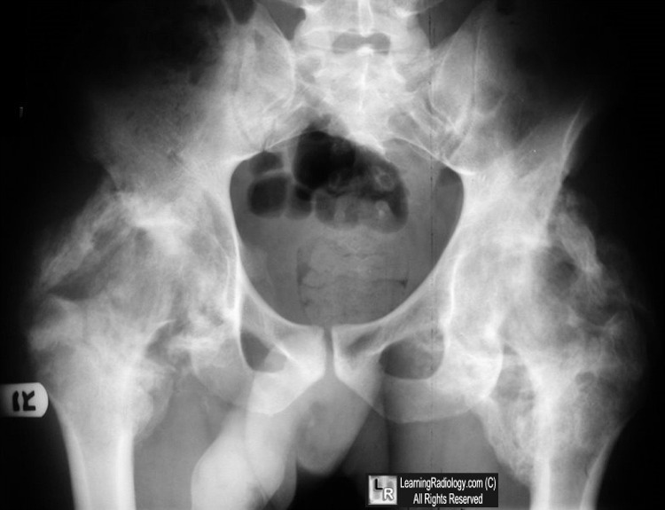
Learningradiology Heterotopic Ossification Ho Myositis Ossificans Radiology Meijer Thanksgiving 2020 Sale
Https Encrypted Tbn0 Gstatic Com Images Q Tbn And9gcsch38ysdfqaqrge8usdwk Yfiqdt Ydj6e Poqcty0btw7lnqk Usqp Cau Meijer Thanksgiving 2020 Sale

Medpix Case Myositis Ossificans Meijer Thanksgiving 2020 Sale
More From Meijer Thanksgiving 2020 Sale
- Safeway Holiday Dinners 2020
- Lunds Holiday Meals
- Queens Thanksgiving Turkey
- Piante Rose Da Giardinohtml
- Cracker Barrel Xmas
Incoming Search Terms:
- Massive Ectopic Soft Tissue Ossifications Is Virtually Diagnostic Of Fibrodysplasia Ossificans Progressiva Fob Formerly Known As M Myositis Radiology X Ray Cracker Barrel Xmas,
- Radiological Identification And Analysis Of Soft Tissue Musculoskeletal Calcifications Insights Into Imaging Full Text Cracker Barrel Xmas,
- Myositis Ossificans Youtube Cracker Barrel Xmas,
- Post Traumatic Myositis Ossificans Cracker Barrel Xmas,
- Myositis Ossificans Portnotes Orthopaedicsone Cracker Barrel Xmas,
- Myositis Ossificans Imaging Keys To Successful Diagnosis Lacout A Jarraya M Marcy Py Thariat J Carlier Ry Indian J Radiol Imaging Cracker Barrel Xmas,